X-Ray Safety and Radiation Leakage:
What every Dental Provider should know
Dental radiography is one of the most valuable tools in modern dentistry, critical for diagnosis, treatment planning, and long-term patient health. The principle behind safe imaging is simple: minimize exposure while preserving diagnostic quality, so the benefits may outweigh the risks.
While exposures to radiation in dental settings are extremely low, ALARA (As Low As Reasonably Achievable) remains the guiding standard for every image captured.
Why X-Ray Safety Matters
X-rays are a form of ionizing radiation. While dental doses are relatively low, repeated exposure without proper precautions subject a user to unnecessary radiation. Safety protocols ensure minimal exposure for patients and maximize protection for clinicians. Ionizing radiation deserves respect, not fear. Every clinician plays a role in maintaining safety for patients and staff through two key practices:
- Justification: Take images only when they can influence diagnosis or treatment planning.
- Optimization: Use the lowest dose that still produces a diagnostically useful image.
Keeping these principles top of mind ensures radiography remains safe and clinically meaningful.
Radiation Leakage vs. Scatter: Not the Same Thing
Leakage and scatter are often used interchangeably, but they describe very different phenomena with distinct safety implications.
Radiation Leakage
Leakage refers to radiation escaping from the X-ray tube in unintended directions. Modern machines have internal shielding around the tube to minimize this. This shielding makes the unit heavier but ensures the safety of the clinician.
Scatter Radiation
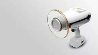
Scatter occurs when X-rays hit an object (like teeth, bone or soft tissue) and change direction. This scattered radiation can bounce back toward the clinician. Proper positioning and shielding are key to minimizing this risk. In addition to the proper positioning of the handheld X-ray, the backscatter shield plays a key role in protecting the clinician.
How to Minimize Exposure
- Position Correctly
- Hold and angle the unit according to manufacturer's instructions.
- Keep the device away from your body; never brace it against yourself.
- Maintain appropriate vertical heights and angles relative to both you and the patient.
- Use the Backscatter Shield Properly
- Position the transparent shield between the source and yourself.
- Ensure it is oriented correctly and not removed or tilted during use.
- Apply Time and Distance Principles
- Use the minimum exposure time necessary for a diagnostic image.
- Whenever possible, increase your distance from the beam; even small increases reduce exposure significantly.
How Shields Work
Backscatter Shields:
Transparent plastics embedded with X-ray–attenuating compounds allow alignment visibility while providing measurable protection from backscatter toward the operator.
Internal Tube Shielding:
X-ray-attenuating compounds also surround the tube housing to contain unintended emissions. This design ensures leakage remains far below regulatory limits and contributes to the overall safety profile of modern handheld and wall-mounted systems.
Dental Radiation in Perspective
Dental imaging contributes only a small fraction of the average person's annual radiation exposure. According to the CDC, medical and dental X-rays use very small amounts of radiation, often comparable to natural background exposure or everyday activities like flying. [cdc.gov]
That perspective underscores a key point: when imaging is justified and performed correctly, it is safe.
Key Takeaways
- Scatter radiation is real, but manageable with proper technique and shielding.
- Radiation leakage occurs at a low rate thanks to built-in shielding, but awareness is essential.
- ALARA remains the guiding principle: justify each image, optimize technique, and position the device, patient and operator correctly.
Dental professionals work at the intersection of precision and safety. Understanding where radiation comes from and how simple habits like proper positioning, and responsible image justification make a difference helps keep exposure minimal without compromising diagnostic excellence.
Safe imaging is not about fear; it is about informed confidence.
Keeping these principles top of mind ensures radiography remains safe and clinically meaningful. The American Dental Association reinforces this approach, emphasizing that X-rays should only be taken when they influence diagnosis or treatment planning, and that the lowest effective dose should always be used.
Contact us today if you want to learn more about the NOMAD Pro 2 and how it’s a safe and effective portable X-ray, perfect for clinicians.
Interested in learning more? Here are some important resources around X-Ray safety:
American Dental Association (ADA) – Overview for X-Rays/Radiographs
- ADA: X-Rays/Radiographs Overview – Covers safety, patient selection, and updated recommendations [ada.org]
- ADA Radiography Safety Update (2024) – New guidance on shielding, beam restriction, and digital imaging [ada.org]
- ADA Radiographic Imaging Guidelines – ALARA principles and patient communication strategies [ada.org]
- ADA Radiation Safety for Pregnant Staff – Workplace safety and accommodations [ada.org]
- ADA/FDA Radiographic Examination Recommendations (PDF) – Detailed guidance on patient selection and exposure limits [ada.org]
CDC – Radiation Safety and Dental Imaging
- Radiation Safety Overview – Covers ALARA principles and protective strategies [cdc.gov]
- Facts About X-Rays – Explains dental X-ray exposure in context [cdc.gov]
- Imaging Procedures and Safety – Details on radiation doses and safety practices [cdc.gov]
FDA – Dental Radiography Guidelines
- Dental Radiography: Doses and Film Speed – Discusses dose reduction through faster film speeds [fda.gov]
- Medical X-ray Imaging Safety – General safety principles for X-ray imaging [fda.gov]
OSHA – Ionizing Radiation Standards
- OSHA Ionizing Radiation Standards – Federal regulations for occupational exposure [osha.gov]
- OSHA Dentistry Safety Overview – Workplace safety considerations for dental professionals [osha.gov]
Quick Safety Checklist for Clinicians
- Use backscatter shield correctly
- Maintain proper distance and angle
- Justify every image
- Follow manufacturer instructions
- Stay informed on local regulations
- Maintain and inspect X-ray units regularly
- Follow ADA and NCRP guidelines
- Keep radiation doses ALARA
DEXIS announces the results of the new independent radiation testing for its handheld, portable X-ray system, NOMAD™ Pro 2. Read the announcement here.


