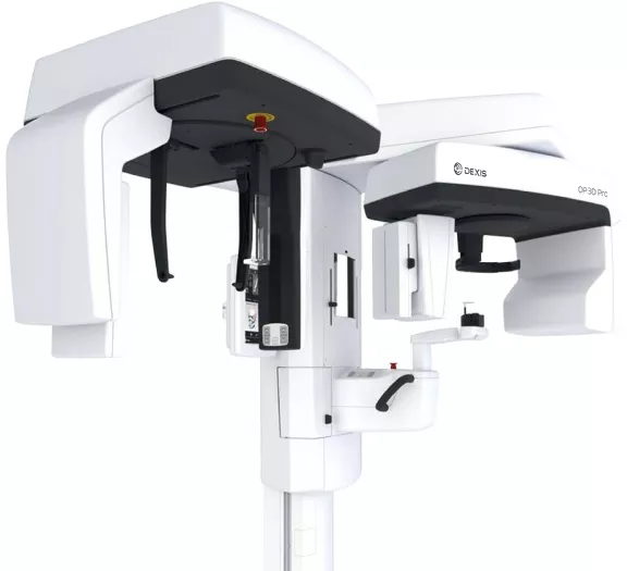
DEXIS™ OP 3D™ Pro
O ORTOHOPANTOMOGRAPH™ 3D Pro da DEXIS™ cristaliza os 50 anos de tradição de uma família lendária de produtos. Ele utiliza o que há de mais recente em tecnologias 3D e 2D para o seu benefício. O design impecável e a versatilidade da plataforma moderna 3 em 1 definem o padrão para a imagem maxilofacial completa.
A solução para todas as suas necessidades: ORTHOPANTOMOGRAPH™ 3D Pro.
O ORTHOPANTOMOGRAPH™ 3D Pro, é uma combinação perfeita da qualidade de imagem de um ORTHOPANTOMOGRAPH com a excelência dos produtos DEXIS™ e máximo conforto operacional. O equipamento oferece imagens de alta precisão em 2D com função de panorâmica multi-camadas e tecnologia de feixe cônico. Além disso, possui quatro resoluções de imagem individuais em 3D, cinco tamanhos de volume, controle automático de dose e a inovadora tecnologia de baixa dose, tornando-o a escolha definitiva para todas as indicações de raios-x, seja usado como um dispositivo 2D padrão ou como um dispositivo 3D, com ou sem opção cefalométrica.
Destaques do DEXIS™ OP 3D™ Pro
Visão geral

Seus benefícios com um DEXIS™ OP 3D™ Pro
- Doses de radiação muito baixas com a Tecnologia de Baixa Dose™.
- Flexibilidade máxima com 5 tamanhos de volume de até FOV 13x15cm e 4 resoluções
- Capacidade de compensar o posicionamento incorreto do paciente e antomias difíceis com o recurso multi-camada, que fornece cinco imagens panorâmicas com apenas uma varredura.
- Obtenção automática da camada de imagem panorâmica mais adequada com o ORTHOfocus™
- Operação simples e intuitiva graças à nova interface do usuário do painel de toque
- Conceito modular que agrega máxima confiabilidade ao investimento.
Clínico geral
Dispositivo de raio-x 3 em 1 para um investimento excelente e confiável.
Endodontistas
Um volume com resolução endodôntica especial e tamanho de volume apropriado para as estruturas mais finas.
Ortodontistas
A mais alta qualidade de imagem para exposições panorâmicas e cefalométricas. Excelente qualidade 3D ajustável para dentes retidos e impactados.
Cirurgia Oral e Maxilofacial
Tamanhos de volume adaptados para toda a região maxilofacial. Funções abrangentes de análise e planejamento no software de raio-x.
Implantodontistas
5 FOVs diferentes com qualidade de imagem otimizada - desde implantes únicos até um conjunto completo, incluindo o planejamento de modelos de perfuração cirúrgica.

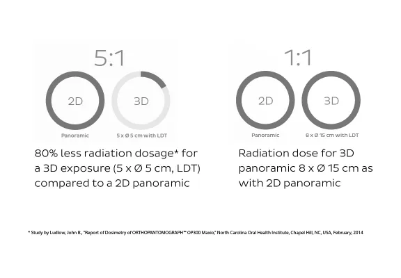
Redução da radiação com a Tecnologia de Baixa Dose™
A inovadora Tecnologia de Baixa Dose™ do DEXIS™ OP 3D™ Pro permite uma qualidade otimizda em imagens de raio-x 3D com uma dose menor de radiação. Para casos sensíveis à dose, em particular, como exposições de acompanhamento ou exposições de crianças, a redução da radiação para proteger seus pacientes representa um valor agregado indispensável.
Adaptação automática da dose de radiação em exposições
ADC para 2D e 3D
A tecnologia ADC otimiza automaticamente os níveis de exposição panorâmica e 3D para cada paciente e cada aquisição, resultando em dosagens específicas para cada paciente e uma eficiência de fluxo de trabalho aprimorada.
ASC
A Compensação Automática da Coluna otimiza a qualidade de imagem através do ajuste da dose ao redor da área da coluna
AFC
O Contorno Facial Automático reduz os fatores de exposição na região dos tecidos moles faciais para proporcionar uma visibiliadde melhorada dos pontos de traçado dos tecidos moles, além de uma redução na dose do paciente.
ORTHOfocus™
Para obeter uma qualidade de imagem panorâmica consistente, o recurso ORTHOfocus™ obtém automaticamente a camada de imagem ideal, permitindo um posicionamento do paciente mais tolerante.
Visão geral
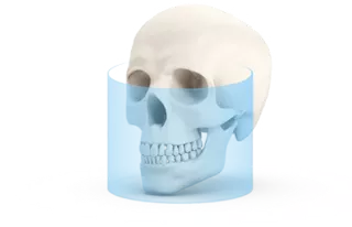
Cinco campos de visão, múltiplas possibilidades.
Para todos os cinco tamanhos de volume, você pode escolher entre três resoluções de imagem. Para o volume de 5 x Ø 5 cm (6 x Ø 4cm*), há uma resolução endo disponível. Cada configuração fornece a resolução perfeita em relação à indicação relevante. Os cinco diferente tamanhos de volume garantem um diagnóstico 3D confiável em toda a região maxilofacial.
Características
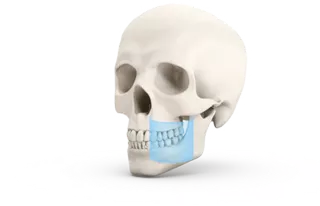
5 x Ø 5 cm (6 x Ø 4 cm*)
Diagnósticos locais:
- Planejamento de implantes individuais;
- Extrações de dentes siso
- Dentes retidos
- Com resolução de endo para imagens de alta precisão das estrutura dos canais e do periodonto.
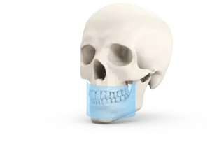
6 x Ø 8 cm
Ilustração de um arco dentário:
Planejamento de múltiplos implantes em uma mandíbula.
Modelos de perfuração
- Na versão de painel pequeno (SFOV) do OP 3D Pro, apenas dois tamanhos de volume 6 x Ø 4 cm e 6 x Ø 8 cm estão disponíveis.
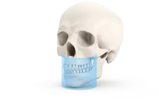
8 x Ø 8 cm
Ilustração de ambas arcadas e partes dos seios maxilares:
- Planejamento de múltiplos implantes e em ambas as mandíbulas;
- Modelos de perfuração;
- Análise de seios maxilares em crianças.
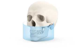
8 x Ø 15 cm
Ilustração da região da mandíbula superior e inferior:
- Ilustração da sinusite maxilar
- Diagnóstico de ATM
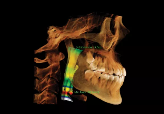
- Coluna vertebral superior e vias respiratórias
- "O 3D panorâmico"
13 x Ø 15 cm*
Ilustração de toda a região maxilofacial:
- Cirurgia maxilar
- Ortodontia
- Diagnóstico de ATM
- Diagnóstico de trauma
- Diagnóstico de Otorrinolaringologia (ENT)
Programas integrados para otimização de qualdiade de imagem
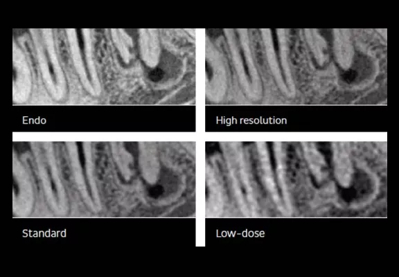
Quatro resoluções
Resoluções individualmente selecionáveis, desde baixa dose até padrão e alta resolução. No volume de 5 x Ø 5 cm (6 x 4 cm*), há também uma resolução endodôntica para imagens de alta precisão das estruturas do canal e do periodonto
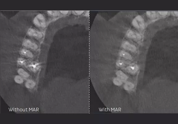
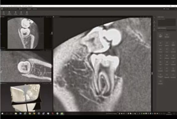
Imagens mais nítidas com a tecnologia MAR
A redução de artefatos metálicos (MAR) selecionável pelo usuário reduz a influência da radiação dispersa, que emerge em estruturas de alta densidade nos volumes de raio-x. Isso otimiza a imagem dos dentes com canais radiculares preenchidos.
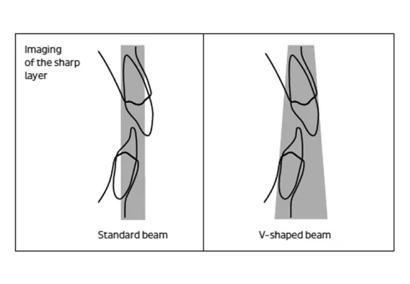
Imagens homogêneas com a tecnologia do feixe cônico
Um feixe em formato cônico considera a melhor absorção da anatomia humanda do que um feixe padrão, garantindo assim uma apresentação de imagem homogênea. Como resultado, as estruturas do maxilar superior são melhor penetradas e a camada nitidamente apresentada no maxilar inferior é significativamente mais ampla.
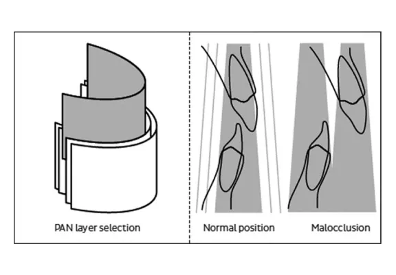
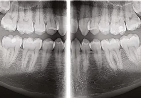
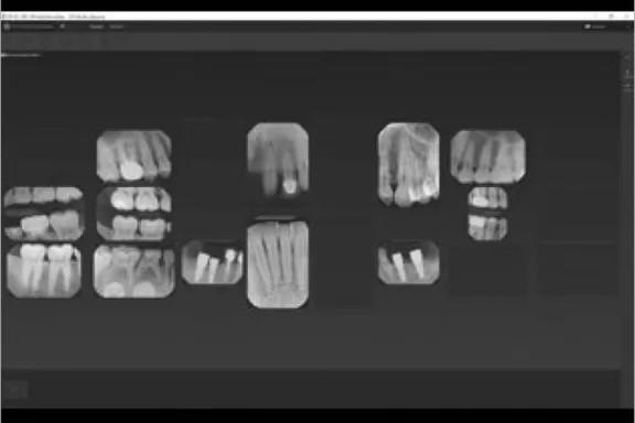
5 é melhor do que 1: função de panorama multicamadas
A função de panorama multicamadas fornece cinco camadas com uma exposição com o mesmo tempo de varrefura e dosagem de uma exposição panorâmica única. A área de foco ampliada através das cinco camadas reduz o risco de novas exposições, por exemplo, em casos de má oclusão.
* Na versão de painel pequeno (SFOV) do OP 3D™ Pro, apenas as duas dimensões de volume 6 x Ø 4 cm e 6 x 8 cm estão disponíveis.
Programas selecionados para diagnósticos excepcionais
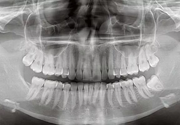
O programa panorâmico padrão fornece definição clara da anatomia dentária, incluindo as ATM. Para crianças, há um programa de colimação de altura e largura para redução da dosagem.
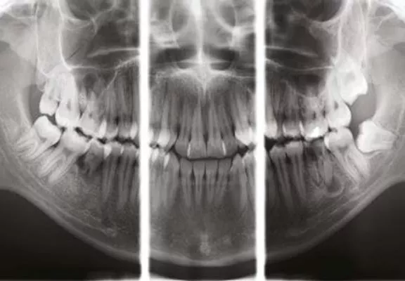
O programa Ortho Zone fornece uma geometria especial com uma camada anterior ampla para pacientes com anormalidades oclusais extremas.
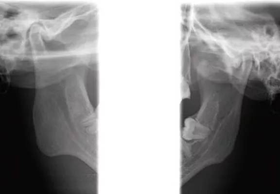
Programas para exposições laterais e frontais da articulação temporomandibular (ATM) com boca aberta ou fechada.
Programa especial para imagens semelhantes e aleta de mordida, com segmentação e colimação específicas.
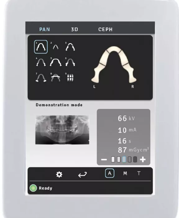
Do fácil ao simplesmente autoexplicativo. O novo painel tátil de 10,4 polegadas.
A operação do DEXIS™ OP 3D™ Pro é projetada para que todos os fluxos de trabalho sejam realizados de forma intuitiva e em questão de segundos. A estrutura clara e os símbolos de fácil compreensão tornam as configurações atuoexplicativas. Seja para exposições 2D ou 3D, o painel tátil de 10,4 polegadas permite uma operação simples e clara, proporcionando confiabilidade operacaional e economia de tempo impressionante.
Com a função SMARTVIEW™, você pode ver antecipadamente o que será registrado posteriormente em 3D
Com a funcionalidade SMARTVIEW™, a precisão de posicioanmento do FOV pode ser verificada ou ajustrda, se necessário, antes do exame CBCT. Além disso, o FOV pode ser posicionado livremente na região de interesse, tanto na direção horizontal quanto vertical com facilidade e confiança.
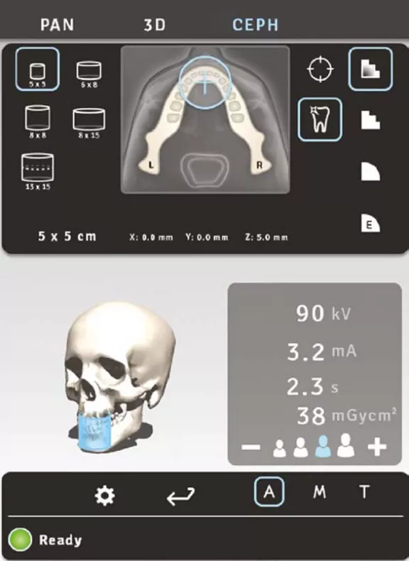
Perfeito, o posicionamento livre do volume na redião de interesse é realizada diretamente pelo painel de toque.
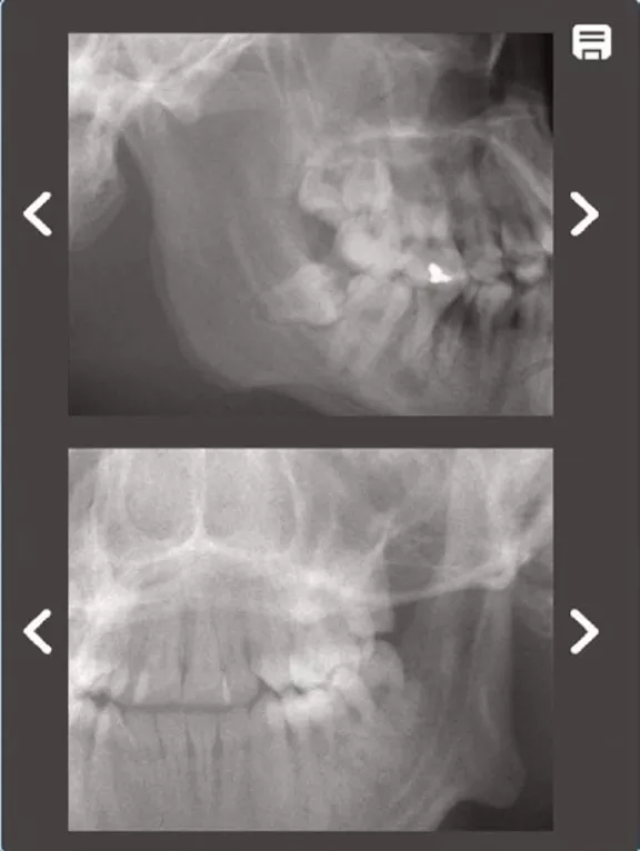
SMARTVIEW™ gera duas imagens prévias em 2D da região sob análise.
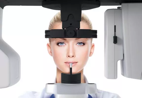
Posicionamento de paciente de 5 pontos para menos artefatos de movimento
Posicione exatamente e matenha essa posição sem desconforto: o posicionamento correto é confirmado por luzes laser de posicionamento operadas automaticamente. Um sistema rígido de posicionamento de 5 pontos reduz o movimento do paciente. O design aberto do produto permite uma visualização e posicionamento fáceis do paciente. O sistema de posicionamento seguro de 5 ponto com apoio de queixo, bloco de mordida e apoio de cabeça, com um ponto de testa e dois pontos de têmporas, reduz o movimento do paciente. Além disso, o design aberto do produto oferece uma visão de primeira classe e permite que você posicione livremente o paciente a partir do lado esquerdo ou direito.
3 em 1 para máxima flexibilidade
O DEXIS™ OP 3D™ Pro é perfeitamente à prova de futuro graças às suas opções de configurações flexíveis. Como um dispositivo panorâmico 2D puro, é ideal para odontologia geral. Além disso, ele pode ser expandido com volumes pequenos/médios ( 6 x Ø4 e 6 x Ø8 cm) ou médios/grandes (5 x Ø5 a 8 x Ø15 ou até 13 x Ø15 cm). Além disso, a opção cefalométrica pode ser posicionada em qualquer um dos lados para um uso ideal do epsaço e uma experiência aprimorada do usuário.
A opção cefalométrica para todas as suas necessidade clínicas
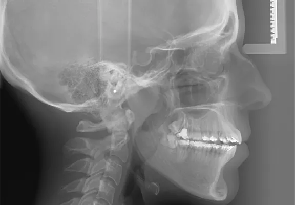
A opção ceflaométrica, que pode ser acoplada ao lado direito ou esquerdo do DEXIS™ OP 3D™ Pro, oferece diversas projeções variadas: lateral cranial, AP/PA, excêntrica cranial e Carpus*. A área de exposição livremente colimável reduz o campo de radiação para a necessidade diagnóstica de cada caso individual.
*Carpus é uma funcionalidade opcional para as unidades DEXIS™ OP 3D™. Entre em contato com o seu representante de vendas local para obter mais informações.
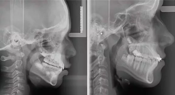
Exposições cefalométricas laterais podem ser geradas em duas alturas diferentes e com colimação de laergura livre entre 17 e 26 cm.
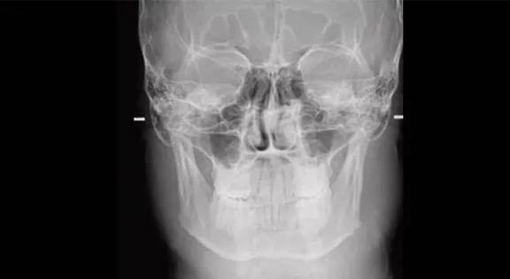
Imagem cefalométrica posterior/anterior. As peças auriculares do aparelho contêm marcações para garantir a posição central.
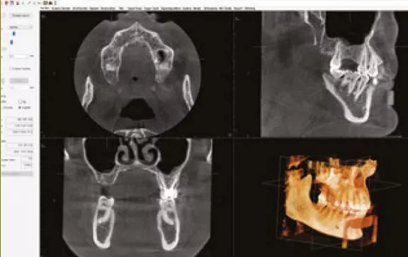
O presente: diagnósticos completos. O futuro: fluxo de trabalho integrado.
O abrangente software de raios-x CLINIVIEW™ será instalado com o seu novo dispositivo. Para imagens em 3D, você pode escolher entre o software de diagnósticos 3D OnDemand™ ou InVitro™. Além disso, você já está preparado para usar a nova plataforma de software unificadora DTX Studio™ para diagnósticos 2D e 3D, abrindo uma nova era de integração de fluxo de trabalho digital.
O software CLINIVIEW™ comprovado e bem conhecido já armazena seus dados compatíveis com a nova plataforma de software DTX Studio™. Seu consultório já estará preparado para aproveitar um fluxo constante de novas melhorias que cobrirão todos os campos da odontologia e tecnologia dentária modernas.
Compatível com os sistemas operacionais Windows e Mac, a plataforma DTX Studio™ integrará tanto os dispositivo existentes e futuros quanto as disposições de software atuais em um processo de trabalho unificado. O CLINIVIEW™ está preparado para suportar uma transição suave para o novo futuro e, passo a passo, abre novas possibilidades que você mesmo não sabia que precisava.
DTX Studio™ fluxo de trabalho uniforme
InVivo™ 3D software de raio-x
DTX Studio™ integração flexível
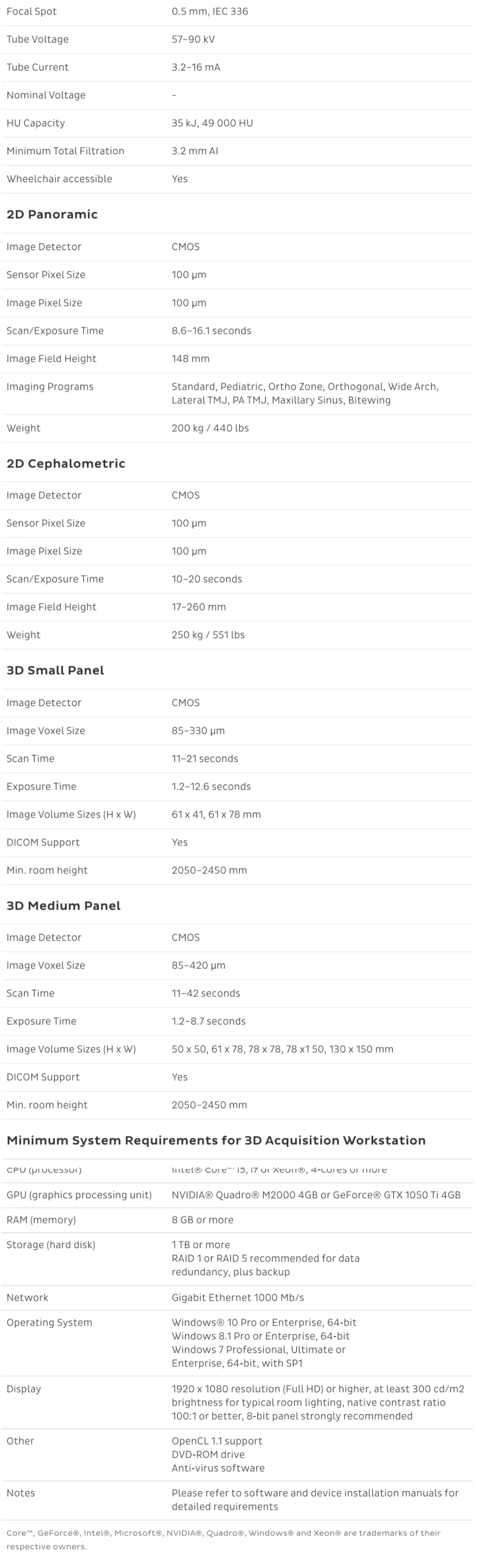
Especificações
To view Documents, please visit the Download Center.