
Long Story Short:
60 Years Innovation
Celebrate our 60-year anniversary with us and explore our legacy of extraoral imaging technology.
Since designing and delivering our very first orthopantomography unit in 1964, our commitment to innovation remains unchanged. Through dedication and innovation, we continue to improve diagnostic quality and diagnostics for the dental industry.
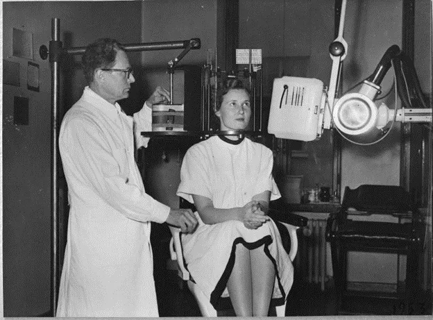
A Pantomograph being used at the University of Helsinki’s Dental Clinic on Fabianinkatu street. Yrjö. V. Paatero on the left. The photo is from an article published in the Uusi Suomi newspaper in 1953.
Photo: Yrjö V. Paatero Archives, privately owned.
A Brief History of X-Ray Imaging Technology
In the world of medical technology, few advancements have had as profound an impact on healthcare as the development of x-ray imaging methods. One of the pioneers in this field was Yrjö Veli Paatero, who revolutionized x-ray imaging techniques.1
The Birth of Parablography
In 1949, Paatero introduced his first x-ray imaging method, which he named parablography. This innovative technique involved placing a strip-like film in the patient's mouth and rotating the patient chair to capture the entire dentition at once. Building upon this initial concept, Paatero further refined his ideas and, in 1950, introduced the Pantomograph™, a breakthrough that allowed for curved layered images of any part of the body.
Paatero's inventive spirit led him to construct the device from parts found in his toolbox. The prototype featured two round plates that could be rotated using a lever, with the x-ray film bent into a U-shape on one plate and the patient's skull placed on the other. This experimental model laid the groundwork for the development of the first panoramic x-ray image of a skull, captured in 1957.1
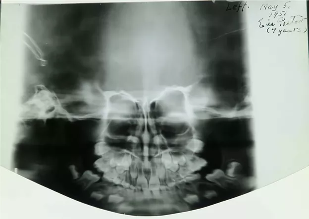
The world’s first panoramic x-ray of a patient’s skull, taken by Yrjö Paatero of his daughter in 1951.
Photo: Yrjö V. Paatero Archives, privately owned.
Recognition and Progress
Paatero's contributions did not go unnoticed. In the 1950s, he received accolades at international invention fairs in Brussels and Paris, and in 1959, he was granted the title of professor by the President of Finland. His expertise in dental radiology led to the establishment of a professorship at the University of Turku, further solidifying his legacy in the field.
As his research continued, Paatero's inventions paved the way for commercial production, with the ORTHOPANTOMOGRAPH™ entering the market in 1961. This marked the beginning of a new era in dental imaging, with Paatero's innovations gaining widespread recognition.1
Evolution Through the Decades
The 1960s and 70s saw significant milestones in x-ray imaging technology, with the founding of Palomex Oy and the commercialization of the ORTHOPANTOMOGRAPH units. The following decades brought forth a wave of advancements, including the introduction of digital imaging and groundbreaking units such as the CRANEX™ Tome – an amalgamation of panoramic and spiral tomographic capabilities.2
In the 2000s, the landscape of x-ray imaging technology continued to evolve. Through mergers and acquisitions, companies like SOREDEX™ and Instrumentarium formed the GE Healthcare Dental P&L, driving further innovations such as the DIGORA™ Optime and the development of 3D dental imaging technology.2
Later in the 2010s, another leap forward with the launch of the first Cone Beam Computed Tomography (CBCT) 3D x-ray device, the SCANORA™ 3D. This milestone cemented the evolution of X-ray imaging into the realm of three-dimensional imaging, offering new possibilities for diagnostics.2
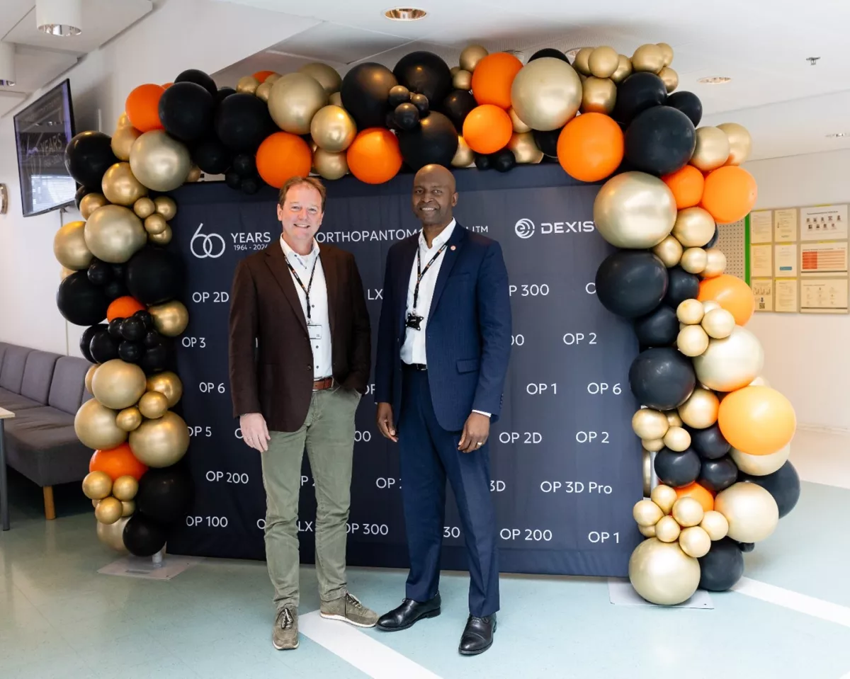
From the left: Stef Vanneste, Senior Director, Europe Commercial, Diagnostics and Robert Befidi, President, Diagnostics
Today, and Looking to the Future
Currently, DEXIS™ continues to play a significant role in advancing dental imaging technology. In 2023, we introduced the DEXIS OP 3D™ LX, an advanced cone beam imaging system that includes 2D and 3D imaging options that cover a full spectrum of dental extraoral needs, from endodontics to complex surgical cases.
Our legacy of continued innovation within the world of dental imaging is far from over. While our 60-year anniversary is a celebration of our past, we’re even more excited to celebrate the future we have planned.


